
90% Less Radiation | Instant Results | Superior Image Quality
Digital X-Rays Austin TX – Advanced Dental Imaging
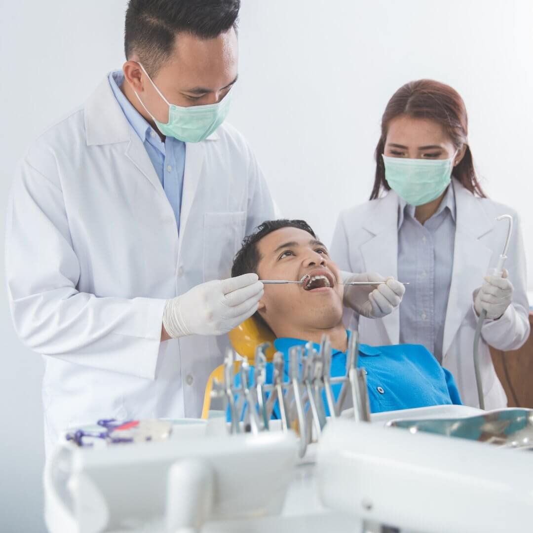
Digital x-rays reveal hidden dental problems using 90% less radiation than traditional film. Instant images help us detect cavities, infections, and bone loss early when treatment is simple and affordable.
See what we see. Modern digital technology provides clearer images, faster diagnosis, and safer radiation exposure. Call +1 (737) 332-4098 to experience advanced dental imaging in Austin TX.
What Are Digital Dental X-Rays?
Digital x-rays use electronic sensors instead of traditional film to capture images of teeth, roots, and surrounding bone. Images appear instantly on a computer screen for immediate diagnosis.
How Digital X-Rays Work:
Traditional film x-rays:
- X-ray beam passes through tissues
- Image captured on film
- Film developed with chemicals (5-10 minutes)
- Single exposure—can’t enhance
- Film stored in physical files
Digital x-rays:
- X-ray beam passes through tissues
- Electronic sensor captures image
- Image appears on screen instantly
- Can be enhanced, enlarged, or adjusted
- Stored electronically (easy retrieval)
The technology is similar to digital cameras replacing film cameras—same concept, better results.
Types of Digital Dental X-Rays
Most common type of dental x-ray. Shows upper and lower teeth in one area of the mouth.
What bitewings reveal:
- Cavities between teeth (invisible during visual exam)
- Bone level between teeth
- Tooth decay under existing fillings
- Tartar buildup below gum line
- Properly fitted crowns and fillings
When taken:
- Routine checkups: Once yearly
- High cavity risk: Every 6 months
- New patients: Initial visit
Number needed: 4 images (right and left, upper and lower)
Duration: 5 minutes total
Bitewings are the workhorse of dental imaging—they catch problems before symptoms appear.
Show entire tooth from crown to root tip plus surrounding bone.
What periapicals reveal:
- Infections at root tips (abscesses)
- Bone loss around roots
- Cysts or tumors
- Impacted teeth
- Root fractures
- Tooth development in children
When taken:
- Tooth pain or swelling
- Before root canal treatment
- After dental trauma
- Evaluating specific problem areas
- New patient full series
Number needed: 1-2 per problem area, or 10-18 for full mouth series
Duration: 1-2 minutes per image
Single image shows entire mouth including all teeth, upper and lower jaws, sinuses, and TMJ joints.
What panoramic x-rays reveal:
- Impacted wisdom teeth
- Jaw fractures or disorders
- Sinus problems
- Bone irregularities
- Tumors or cysts
- TMJ conditions
- Overall tooth development
When taken:
- New patients
- Wisdom tooth evaluation
- Orthodontic planning
- Implant planning
- TMJ assessment
- Every 3-5 years for updates
Number needed: 1 image captures everything
Duration: 2-3 minutes
You stand or sit while machine rotates around your head—completely painless.
Three-dimensional imaging provides detailed views impossible with traditional x-rays.
What 3D scans reveal:
- Exact bone dimensions for implant placement
- Root canal anatomy (extra canals, curves)
- Impacted tooth position
- Jaw fractures (exact location)
- Airway obstructions
- Sinus anatomy
- TMJ detailed structure
When needed:
- Dental implant planning (most common)
- Complex root canals
- Surgical extractions
- Jaw surgery planning
- TMJ evaluation
- Orthodontic assessment
Number needed: 1 scan captures entire area
Duration: 10-20 seconds of scanning, 5 minutes total
Cost: $300-$500 (insurance may cover for medical necessity)
3D imaging has revolutionized implant dentistry—we see exactly what we’re working with before surgery begins.
Benefits of Digital X-Rays
90% Less Radiation Exposure
Digital sensors require far less radiation to produce images compared to film.
Radiation comparison:
- Traditional film x-ray: 0.005 mSv
- Digital x-ray: 0.0005 mSv (90% reduction)
- Daily natural background radiation: 0.01 mSv
- Airplane flight (cross-country): 0.03 mSv
- Chest x-ray: 0.1 mSv
Digital x-rays expose you to less radiation than:
- One day of natural background radiation
- Two hours on an airplane
- One hour in the sun
Safety measures we use:
- Lead apron (protects body)
- Thyroid collar (protects thyroid)
- Collimated beam (targets only necessary area)
- Minimal exposure time
- Only necessary x-rays (ALARA principle)
Digital x-rays are safe for:
- Children
- Adults
- Seniors
- Pregnant women (when necessary with precautions)
Instant Image Availability
No waiting for film development.
Traditional film:
- Exposure: 1 minute
- Development: 5-10 minutes
- Fixing and drying: 5 minutes
- Total time: 10-15 minutes
Digital x-rays:
- Exposure: 1 minute
- Image appears: Instantly
- Total time: 1 minute
Benefits of instant imaging:
- Faster diagnosis
- Same-day treatment planning
- Immediate patient education
- Retake bad images immediately (rare)
- No waiting in chair
We can show you problems immediately—you see exactly what we see.
Superior Image Quality
Digital images are clearer and more detailed than film x-rays.
Enhancement capabilities:
- Zoom in on specific areas
- Adjust brightness and contrast
- Highlight problem areas with color
- Measure distances precisely
- Compare current and previous images side-by-side
Better diagnosis through:
- Enhanced cavity detection
- Clearer bone loss visibility
- Improved infection identification
- Precise measurement tools
- Superior detail in root canals
We can enlarge images 10-20 times—tiny cavities become clearly visible.
Environmentally Friendly
Digital x-rays eliminate toxic chemicals used in film development.
Traditional film processing requires:
- Developer chemicals (toxic)
- Fixer chemicals (toxic)
- Wash water (contaminated)
- Disposal of hazardous waste
- Lead foil packets (hazardous)
Digital x-rays use:
- Electronic sensors (reusable)
- No chemicals
- No contaminated water
- No hazardous waste
- Minimal environmental impact
Going digital means:
- Cleaner environment
- No toxic chemical exposure for staff
- Reduced water consumption
- Less hazardous waste
Easy Storage and Retrieval
Digital images stored electronically never degrade or get lost.
Traditional film:
- Physical storage required
- Images degrade over time
- Can be lost or damaged
- Difficult to share with specialists
- Takes time to retrieve
Digital storage:
- Unlimited cloud storage
- Images never degrade
- Cannot be lost
- Instant sharing with specialists
- Retrieve in seconds
- Compare with previous images easily
We can send your x-rays to specialists electronically in minutes—no waiting for film copies.
Better Patient Education
Seeing is believing.
Digital x-rays help patients understand:
- Exact location of cavities
- Severity of gum disease (bone loss visible)
- Why treatment is recommended
- How problems developed
- Progress of treatment
We display images on large screens:
- Point to specific problems
- Show before and after comparisons
- Zoom in on areas of concern
- Use color highlighting
- Print images for your records
Patients who see their x-rays:
- Better understand treatment needs
- More likely to complete treatment
- Less likely to delay necessary care
- More engaged in their oral health
What Digital X-Rays Detect
Cavities Between Teeth
40% of cavities form between teeth where visual examination and probing can't reach.
Bitewing x-rays reveal:
- Early decay (can be reversed with fluoride)
- Small cavities needing fillings
- Large cavities approaching nerves
- Decay under old fillings
Early detection saves:
- Tooth structure (smaller fillings)
- Money ($150 filling vs. $2,500 root canal + crown)
- Pain (treat before nerve involvement)
Bone Loss from Gum Disease
Gum disease destroys bone supporting teeth.
X-rays show:
- Current bone level
- Amount of bone loss
- Pattern of bone loss
- Prognosis for affected teeth
Early detection allows:
- Deep cleaning to stop progression
- Bone grafting (in some cases)
- Tooth preservation
Without x-rays, bone loss goes unnoticed until teeth become loose.
Gum disease treatment →
Infections at Root Tips (Abscesses)
Tooth infections appear as dark areas around root tips on x-rays.
Abscesses indicate:
- Dead or dying tooth nerve
- Bacterial infection in bone
- Need for root canal or extraction
Detecting infections early:
- Prevents spread to other teeth
- Avoids facial swelling
- Reduces risk of systemic infection
- Allows less invasive treatment
Some infections cause no pain initially—x-rays catch them before symptoms develop.
Tooth infection treatment →
Impacted Wisdom Teeth
Panoramic x-rays show wisdom tooth position and potential problems.
X-rays reveal:
- Impaction angle
- Proximity to nerves
- Damage to adjacent teeth
- Cyst formation
- Infection risk
Early evaluation allows:
- Planned extraction (not emergency)
- Easier removal (before roots fully form)
- Prevention of damage to second molars
Most wisdom teeth should be removed between ages 17-25 when risks are lowest.
Bone Irregularities and Tumors
Abnormal bone growths appear on x-rays.
X-rays detect:
- Benign cysts
- Bone spurs
- Jaw tumors
- Bone infections (osteomyelitis)
Early detection is critical:
- Benign tumors treated before becoming malignant
- Infections caught before extensive damage
- Surgical planning for removal
Developmental Issues in Children
Children's x-rays show tooth development.
X-rays reveal:
- Missing permanent teeth
- Extra teeth
- Abnormally shaped teeth
- Improper tooth positioning
- Development timing
Early detection allows:
- Orthodontic planning
- Timely intervention
- Prevention of future problems
Digital X-Ray Frequency
How Often You Need X-Rays:
Low cavity risk:
- Bitewings: Every 18-24 months
- Full series: Every 3-5 years
- Panoramic: Every 3-5 years
Moderate cavity risk:
- Bitewings: Every 12 months
- Full series: Every 3 years
- Panoramic: Every 3 years
High cavity risk:
- Bitewings: Every 6 months
- Full series: Every 18-24 months
- Panoramic: As needed
Gum disease patients:
- Bitewings: Every 6-12 months
- Full series: Every 2-3 years
- Panoramic: As needed
New patients:
- Full series or panoramic: Initial visit
- Establishes baseline
We take only necessary x-rays following ALARA guidelines (As Low As Reasonably Achievable).
Digital X-Ray Safety
Minimal Radiation Exposure
Digital dental x-rays are among the safest medical imaging procedures.
Radiation dose comparison:
- Full mouth digital x-rays: 0.009 mSv
- One day natural background: 0.01 mSv
- Cross-country flight: 0.03 mSv
- Chest x-ray: 0.1 mSv
- CT scan of abdomen: 10 mSv
Annual radiation limits:
- General public: 1 mSv per year (x-rays are 1% of this)
- Radiation workers: 50 mSv per year
Your dental x-rays represent less than 1% of safe annual exposure.
Protection Measures
We use multiple safety precautions:
Lead apron:
- Protects body organs
- Blocks 95-99% of scatter radiation
- Worn during all x-rays
Thyroid collar:
- Protects thyroid gland
- Especially important for children
- Standard for all patients
Collimated beam:
- Targets only area being imaged
- Minimizes radiation exposure
- Reduces scatter radiation
Fast sensors:
- Require minimal exposure time
- High sensitivity
- Instant imaging
Trained staff:
- Proper positioning (first time)
- Minimal retakes
- ALARA principle followed
Pregnancy and X-Rays
We avoid x-rays during pregnancy when possible.
When x-rays are necessary:
- Emergency situations
- Infection diagnosis
- Trauma evaluation
Safety measures for pregnant patients:
- Double lead apron
- Thyroid collar
- Minimal number of images
- Digital technology (lowest radiation)
Dental x-rays pose minimal risk to developing babies when proper precautions are used.
Untreated dental infections pose greater risk than diagnostic x-rays.
Digital X-Ray Cost in Austin TX
X-Ray Pricing:
Bitewing x-rays (4 images):
- Cost: $50-$100
- Insurance covers: 80-100%
- Your cost: $0-$20
Full mouth series (18 images):
- Cost: $150-$250
- Insurance covers: 80-100%
- Your cost: $0-$50
Panoramic x-ray:
- Cost: $100-$150
- Insurance covers: 80-100%
- Your cost: $0-$30
3D cone beam CT:
- Cost: $300-$500
- Insurance covers: Variable (if medically necessary)
- Your cost: $100-$500
Insurance Coverage:
Most dental insurance covers:
- Bitewings: Once yearly (100%)
- Full series: Every 3-5 years (100%)
- Panoramic: Every 3-5 years (80-100%)
We verify benefits before taking x-rays and explain any costs.
Digital X-Ray – FAQs
Yes, digital x-rays are extremely safe. They use 90% less radiation than film x-rays and expose you to less radiation than one day of natural background radiation or two hours on an airplane. We use lead aprons, thyroid collars, and follow ALARA principles (As Low As Reasonably Achievable) for maximum safety.
Frequency depends on your cavity and gum disease risk:
- Low risk: Bitewings every 18-24 months
- Moderate risk: Bitewings every 12 months
- High risk: Bitewings every 6 months
- Full series: Every 3-5 years for updates
We take only necessary x-rays based on your individual needs.
We avoid x-rays during pregnancy unless absolutely necessary for emergency diagnosis or treatment. When required, we use double lead aprons, thyroid collars, and minimal images. Digital x-rays pose minimal risk to developing babies. Untreated dental infections pose greater risk than diagnostic x-rays.
Digital x-rays use electronic sensors instead of film:
- 90% less radiation exposure
- Instant results (no film development)
- Superior image quality
- Can be enhanced and enlarged
- Environmentally friendly (no chemicals)
- Easy storage and sharing
- Better patient education
Results are identical or better with significantly improved safety.
No, x-rays are completely painless. You'll feel:
- Plastic sensor placed in mouth (firm but not painful)
- Brief pressure holding sensor in place
- Nothing during actual x-ray (1-2 seconds)
Some patients find sensors uncomfortable due to size, but improved sensor designs minimize discomfort.
X-rays detect 40% of cavities that visual exams miss (between teeth). However:
- Cavities on smooth surfaces may not show on x-rays
- Very early decay might not be visible yet
- Some angles hide small cavities
We use x-rays combined with visual examination and probing for complete cavity detection.
Most dental problems cause no symptoms until advanced:
- Cavities don't hurt until reaching the nerve
- Gum disease is painless until severe bone loss
- Infections can be present without pain
- Tumors and cysts develop silently
X-rays detect problems early when treatment is simple, less expensive, and less painful. Waiting for pain means waiting too long.
X-ray duration varies by type:
- Bitewings (4 images): 5 minutes
- Full series (18 images): 15-20 minutes
- Panoramic: 2-3 minutes
- 3D cone beam CT: 10-20 seconds scanning, 5 minutes total
Most routine appointments include 4 bitewings taking about 5 minutes.
Yes, x-rays require your consent. However, refusing x-rays:
- Prevents complete diagnosis
- May delay or prevent treatment
- Could result in undiagnosed problems
- Limits our ability to provide care
We explain why x-rays are necessary and address any concerns. Informed patients rarely refuse recommended x-rays.
We can request x-rays from your previous dentist electronically. Bring this information:
- Previous dentist name and phone number
- Approximate date of x-rays
We'll transfer recent x-rays to avoid unnecessary duplication. However, if previous x-rays are over 12 months old or don't show needed areas, new x-rays may be necessary.
Experience Modern Dental Imaging
Digital x-rays provide safer, faster, and more accurate diagnosis than traditional film. Early detection prevents pain, saves teeth, and costs far less than treating advanced problems.
See the difference digital technology makes.